This Changes Everything About Predicting Heart Attacks
In a Nutshell
Coronary CT angiography with AI analysis detects dangerous soft plaques and arterial inflammation that calcium scans and stress tests miss, predicting heart attacks far more accurately than current methods. Standard cholesterol tests, risk calculators, stress tests, and calcium scores fail because they don't measure the unstable plaques that actually cause heart attacks, while this scan correctly identifies high-risk patients and enables targeted treatment. Insulin resistance drives most plaque formation, so testing insulin levels and inflammation alongside the scan provides the complete picture needed for prevention.
These notes were generated by AI and may contain inaccuracies.
The way we predict heart attacks is about to change forever. For people over fifty worried about heart attacks, the standard approach involves stress tests, calcium level checks, cholesterol testing, and statin prescriptions, followed by hoping things go well. This approach is the reason heart attacks remain the number one cause of death worldwide because doctors cannot predict when a heart attack will occur and do not know how to prevent them.
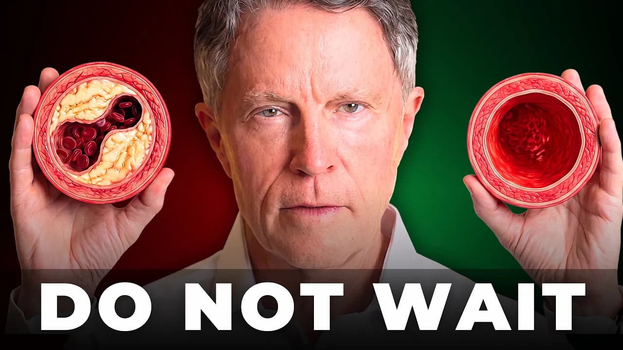
A completely new study shows one test detects arterial plaques better than any other test. This test has been used for several years and is the gold standard. It detects not just stable calcified plaques found by calcium scans, but also soft, unstable, and dangerous plaques that can cause heart attacks. Stable plaques pose a risk almost equal to having no plaques at all and will not cause a heart attack.
The current approach to preventing heart attacks is flawed in four key areas, each either incomplete or a complete deception. First, cholesterol numbers and immediate statin prescriptions neglect metabolic diseases while focusing on cholesterol, leading to more health incidents despite LDL cholesterol and apoB playing a role in cardiovascular risk. Second, the Framingham risk calculator only counts metabolic disease when overt diabetes has already been diagnosed, missing the years of risk from insulin resistance and prediabetes before diabetes is diagnosed.
Stress testing cannot predict heart attacks because it only detects blockages cutting off more than 50% of blood flow. The COURAGE, ORBITA, and ISCHEMIA experiments all showed this approach is ineffective. The PROMISE experiment with over 9,000 patients found more than half of heart attacks and deaths came from people without arterial blockages. The ISCHEMIA experiment with 5,000 patients who tested positive on stress tests showed no difference in deaths or heart attacks between those receiving stents/bypass surgery versus medication alone. The severity of blockage did not predict who would die.
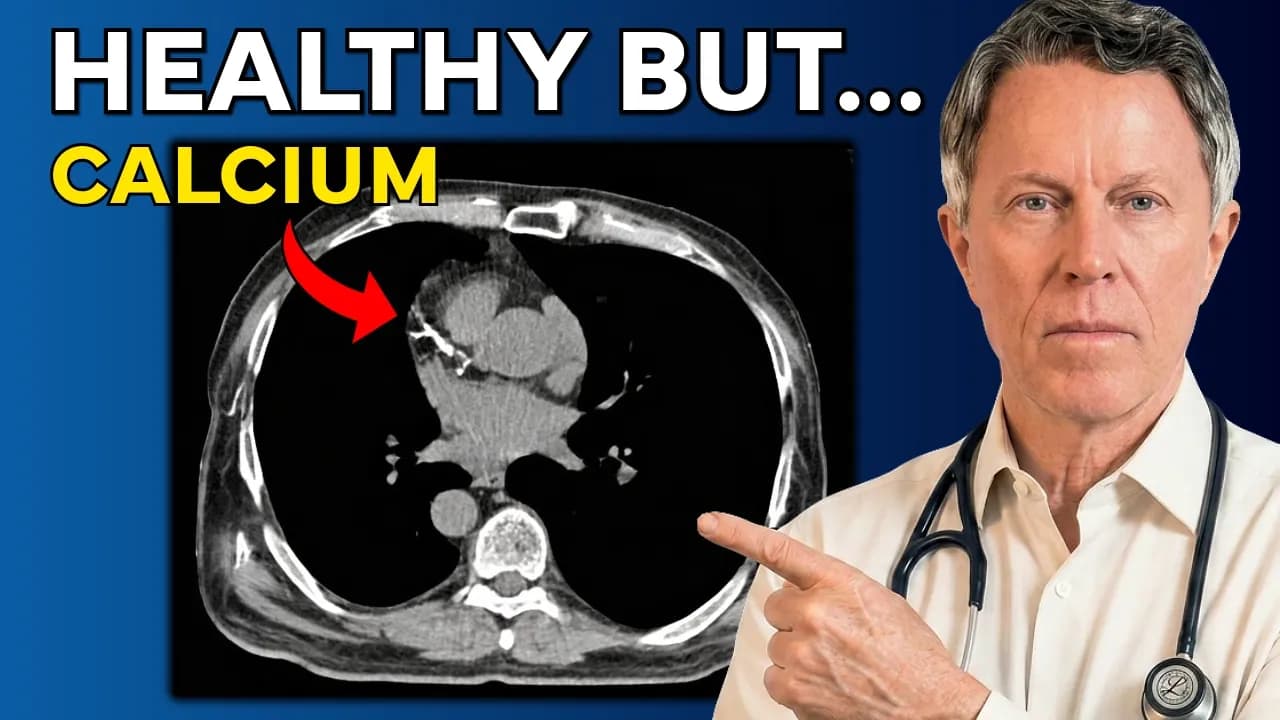
Calcium scores are better than stress tests at predicting risk, but the main drawback is they cannot find soft, unstable, and inflamed plaques. They only reveal stable, calcified plaques. Previously, when people did not address metabolic diseases, this logic had credibility. Now that people are changing lifestyles and stopping new plaque accumulation, the numbers have changed dramatically. A positive calcium test means plaques exist and vascular disease is present, but interpretation based on low versus high results is no longer realistic.
The SCOT-HEART trial showed 13% of people with zero calcium scores had more than 4% of their artery volume filled with soft plaques. One in 10 heart attacks in the trial occurred in people with zero calcium scores. The American Heart Association agreed five years ago that if you don't have plaques, you don't need statins, but they measured plaque absence using calcium scores, which is incorrect. Calcium scoring counts scars that no longer pose a threat but does not count the current threat.
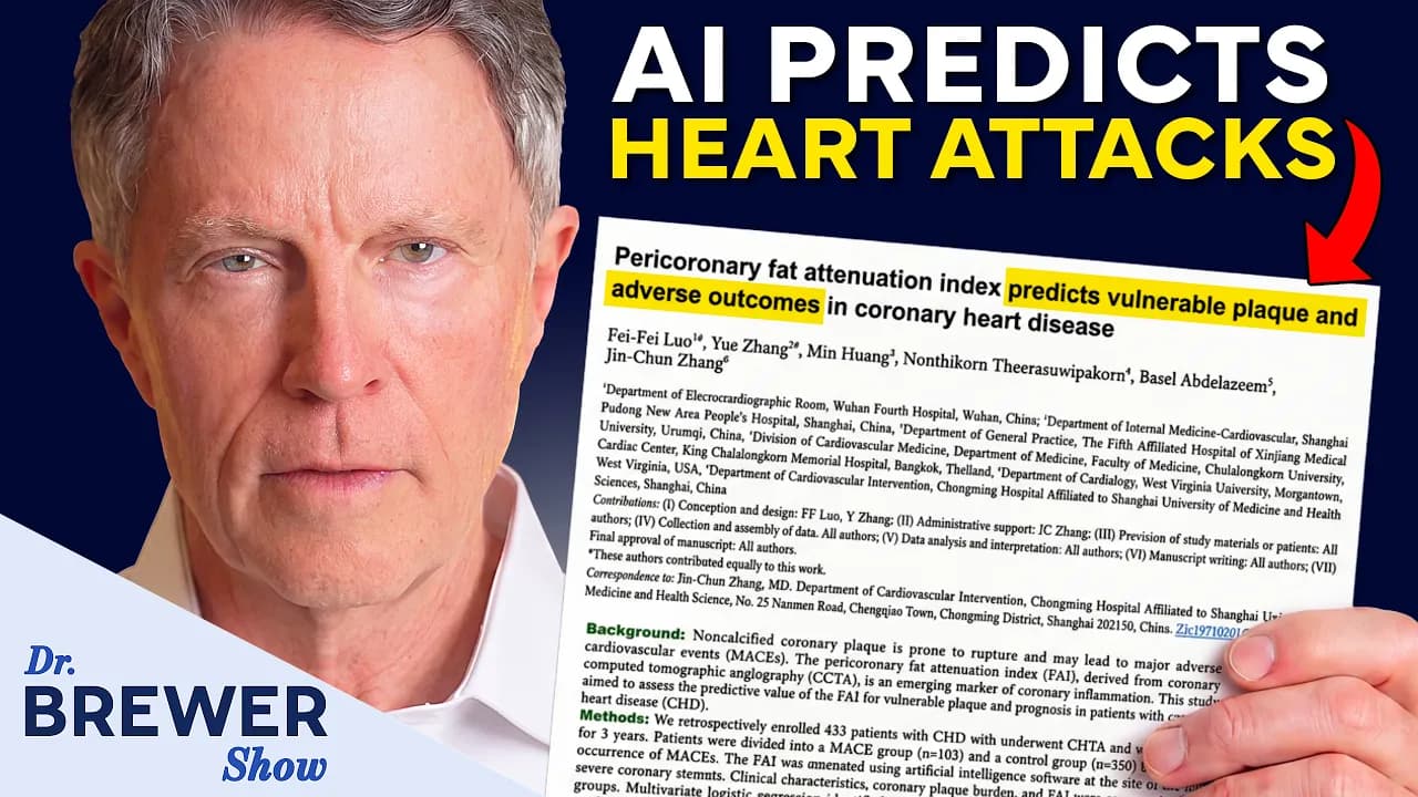
A heart attack results from soft deposits and an inflamed artery wall. None of the four current elements (cholesterol tests, risk calculators, stress tests, calcium scoring) assess this risk. The test that reveals this risk is a tomography scan of the coronary arteries with deposit analysis using artificial intelligence, known as coronary computed tomography angiography with AI analysis.
Coronary computed tomography angiography has existed for over a decade, but the new element is the AI analysis software. The human eye cannot see these deposit levels or distinguish soft deposit amounts. The software measures every millimeter of deposits in each coronary artery and classifies them as calcified, non-calcified, or low-density soft deposits (the type that tears).
The CONFIRM 2 trial published this year followed 3,500 patients for 4 years. The software measured 24 items per scan, but only two predicted heart attacks independently: the amount of non-calcified deposits in the wall and how narrow the artery is. The strongest single indicator was soft, non-calcified plaques. Using usual risk factors alone correctly predicts heart attacks six times out of ten. Adding plaque numbers from the scan improves this to three times out of four, correctly predicting 15 additional heart attacks per 100 pairs.
The SCOT-HEART trial followed 1,700 patients for approximately 5 years. When soft plaques comprised more than 4% of artery volume, heart attack risk increased four and a half times. Calcium levels had almost no effect on results once soft plaques were included. The stress test findings were not statistically significant once soft plaques were included. This trial was a randomized clinical trial where doctors treated what they saw with more preventative treatment and the same number of cardiac catheterizations, resulting in a 41% decrease in heart attacks and coronary artery deaths.
The PROMISE trial used the same footage without instructions to work based on it, and nothing changed. This demonstrates that measuring deposits has no value unless someone treats those deposits. The ISCHEMIA group observed the same thing when applying this software to their 3,700 scans: once deposit size was entered into the model, the number of blocked arteries ceased to be important.
Soft plaques are only half of what this test measures. The other half is inflammation, which leads to soft plaque rupture. Each coronary artery is covered with a layer of fat. When the artery wall beneath becomes inflamed, it sends chemical signals preventing nearby fat cells from storing fat. In CT scans, the density of that fat changes, and the computer can measure this change. The fat surrounding the artery becomes an indicator of inflammation within that artery.
The ORPHAN study published in The Lancet in 2024 included 40,000 people who underwent computed tomography angiography. 81% did not have obstructive disease but represented two-thirds of heart attack cases and deaths. For people with severe inflammation around the three coronary arteries, the risk of cardiac death was about 30 times higher. The Princeton Prevention Institute has stated the vast majority of heart attacks, strokes, and deaths occur in people with no effect on blood flow.
This test requires a doctor order because it is expensive and cannot be ordered directly. It is a CT scan of the heart using contrast dye with an intravenous catheter. Beta-blockers are usually taken to slow heart rate for clearer images. The examination takes a few seconds while holding breath, with the whole process taking less than 20 minutes and definitely less than an hour. The sediment analysis is performed by AI and the report returns to the doctor within a day or two.
The test costs approximately $2,000 out of pocket for asymptomatic people. As of January 2026, AI-powered sediment analysis will have its own permanent billing token. Medicare pays about $1,000 for the analysis plus a few hundred dollars for the test itself, but only if symptoms are present. Insurance coverage currently excludes asymptomatic people from preventive screenings. Aetna, United Healthcare, Cigna, and Humana have started covering costs for eligible patients. One San Diego imaging center advertises $1,950 cash price for the scan with AI plaque analysis.
There is radiation involved, though much less than previously and close to zero compared to heart attack and stroke risks. The test requires preparation because heart rate needs to drop for clear images. Many patients pay out of pocket despite not being wealthy, prioritizing this test over new vehicles, boats, jet skis, cruises, or expensive golf. The $2,000 cost equals less than three new car payments, about four used car payments, or less than one person's share of a typical summer vacation.
As of January, AI-powered plaque analysis has its own permanent billing token, which is how medicine determines something is real enough to pay for. Medicare now pays around $1,000 for this analysis. Coverage is moving in the right direction, though currently only for people meeting specific criteria. Most people still pay cash, but the payment landscape is improving.
Comparing the expense of advanced testing to other lifestyle purchases like a truck, trip, vacation, jet ski, or boat helps frame the decision. The speaker chose to keep driving their Lexus with 120,000 miles rather than spend on new vehicles, determining that the testing was worth the trade-offs. This represents the best current approach for answering critical health questions.
After finding plaques through testing, the immediate priority is understanding why they formed. The first step of the protocol is to test, don't guess. This involves determining whether arteries are still being damaged internally and identifying the root causes of plaque development.
The protocol requires measuring insulin resistance and inflammation in the body. An oral glucose tolerance test that includes insulin measurement is essential, as relying solely on glucose readings will miss 20% of cases. A continuous glucose monitoring device should be used for two weeks, which doesn't require a prescription.
The triglyceride to HDL ratio provides valuable information and can be obtained from various laboratories. Inflammation assessment requires going beyond standard C-reactive protein to include markers that indicate whether plaques are active within vessels, such as MPO (myeloperoxidase), LP-PLA2, and urine ratio tests.
Fasting glucose and A1C tests are insufficient because they become positive years after plaques begin forming. The body requires elevated insulin levels to maintain glucose control, allowing plaques to develop over years or decades while glucose levels remain normal. The American Diabetes Association acknowledges this in their research but continues to recommend only these standard tests, which address less than half of the problem.
Insulin resistance is identified as the root cause in more than 90% of plaque buildup, heart attacks, and strokes cases. This is the test most doctors don't order, as finding plaques is only the first step—determining why they exist is the crucial next step.
The recommended approach begins with imaging that detects soft plaques, followed by insulin and inflammation testing to identify triggers, then building personalized plans based on individual numbers. This differs from standard care that focuses only on monitoring LDL cholesterol and ordering routine stress tests.
For those unable to access radiation-based testing, another non-radiation test exists for finding soft plaques, though finding quality versions of this test can be challenging.
Getting old is not for cowards.
At age sixty-nine, the speaker notes that maintaining healthy arteries makes aging significantly easier.
Keep Dr. Ford Brewer MD MPH in your library
Save the videos and channels worth coming back to, and find them again in one place.